Facebook Youtube Instagram Contacts e-mail Phone
Doctor's phone: +49 681 406-4692
Make an appointment: +49 681 406-1370
Surgical management of uterine leiomyoma

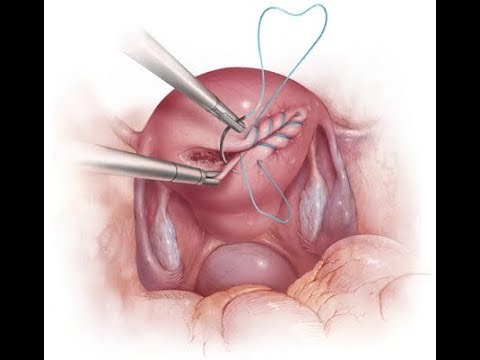
Surgical treatment for uterine leiomyoma is divided into two primary categories: organ-preserving procedures (myomectomy) and radical surgery (hysterectomy).

Surgical indications: Dominant fibroid > 5–6 cm, rapid tumor growth (doubling within a year or less), total uterine volume exceeding 12 weeks of gestation, and severe clinical manifestations (abnormal uterine bleeding, anemia, chronic pelvic pain, compression of adjacent organs).

Uterine preservation is determined individually with each patient. To ensure bloodless, precise myomectomy, we routinely perform temporary clipping of the ascending uterine arteries. In patients with completed childbearing or concurrent cervical/endometrial pathology, laparoscopic subtotal (LASH) or total intrafascial hysterectomy (TLH) is performed.


I. Organ-preserving surgery (Laparoscopic myomectomy)


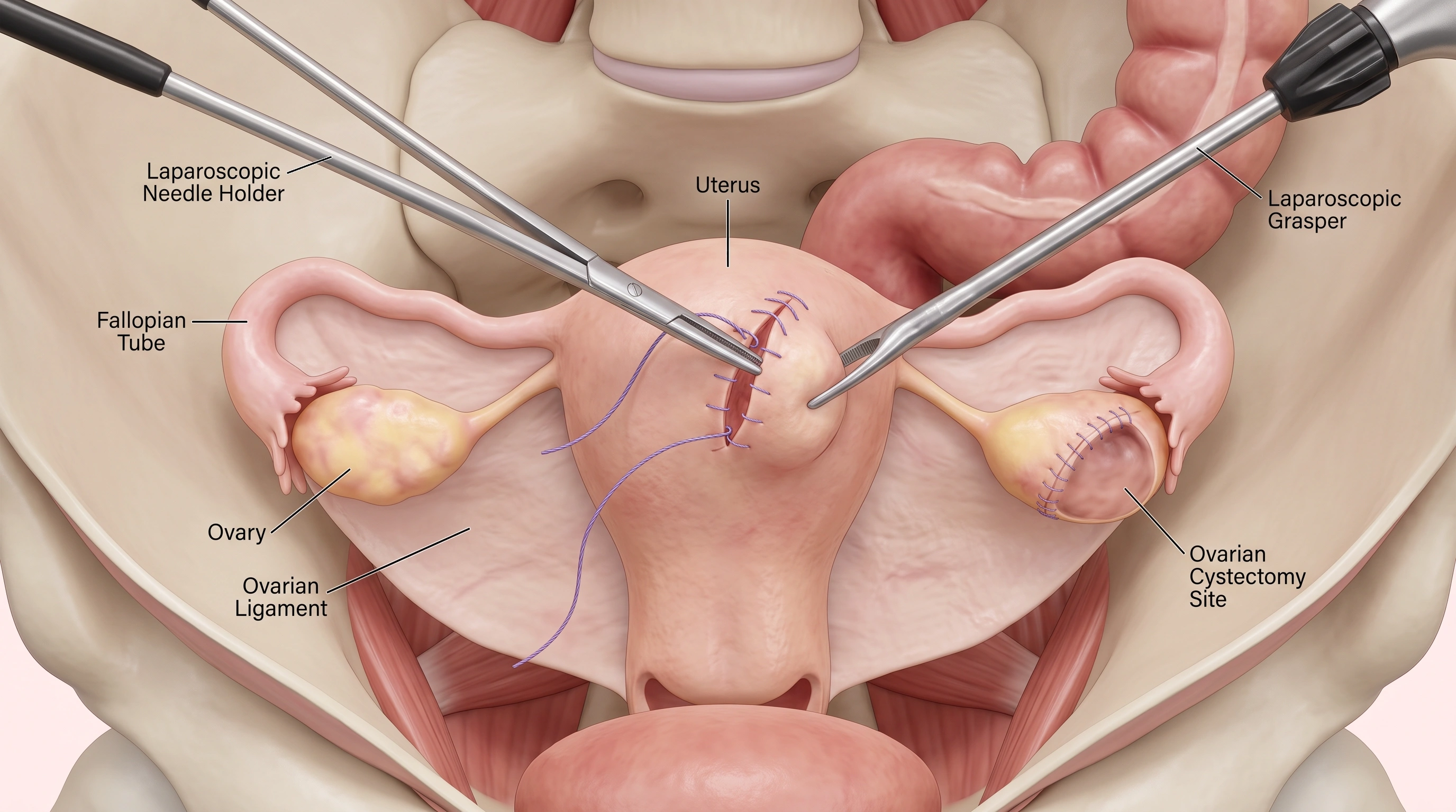
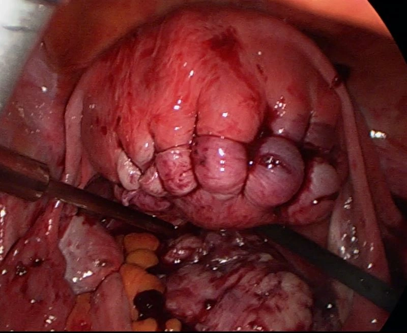
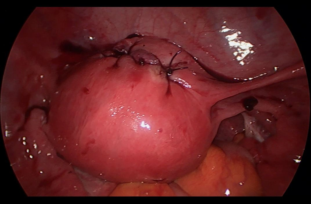
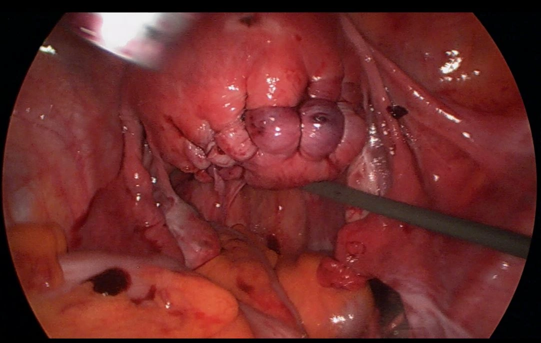
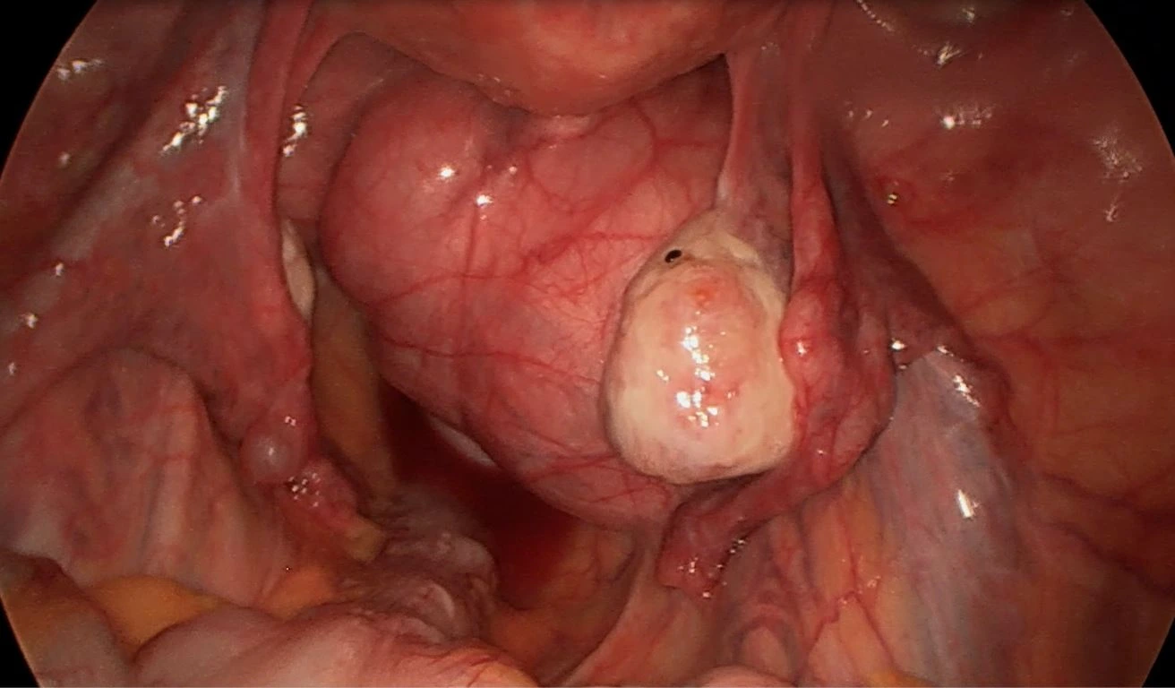
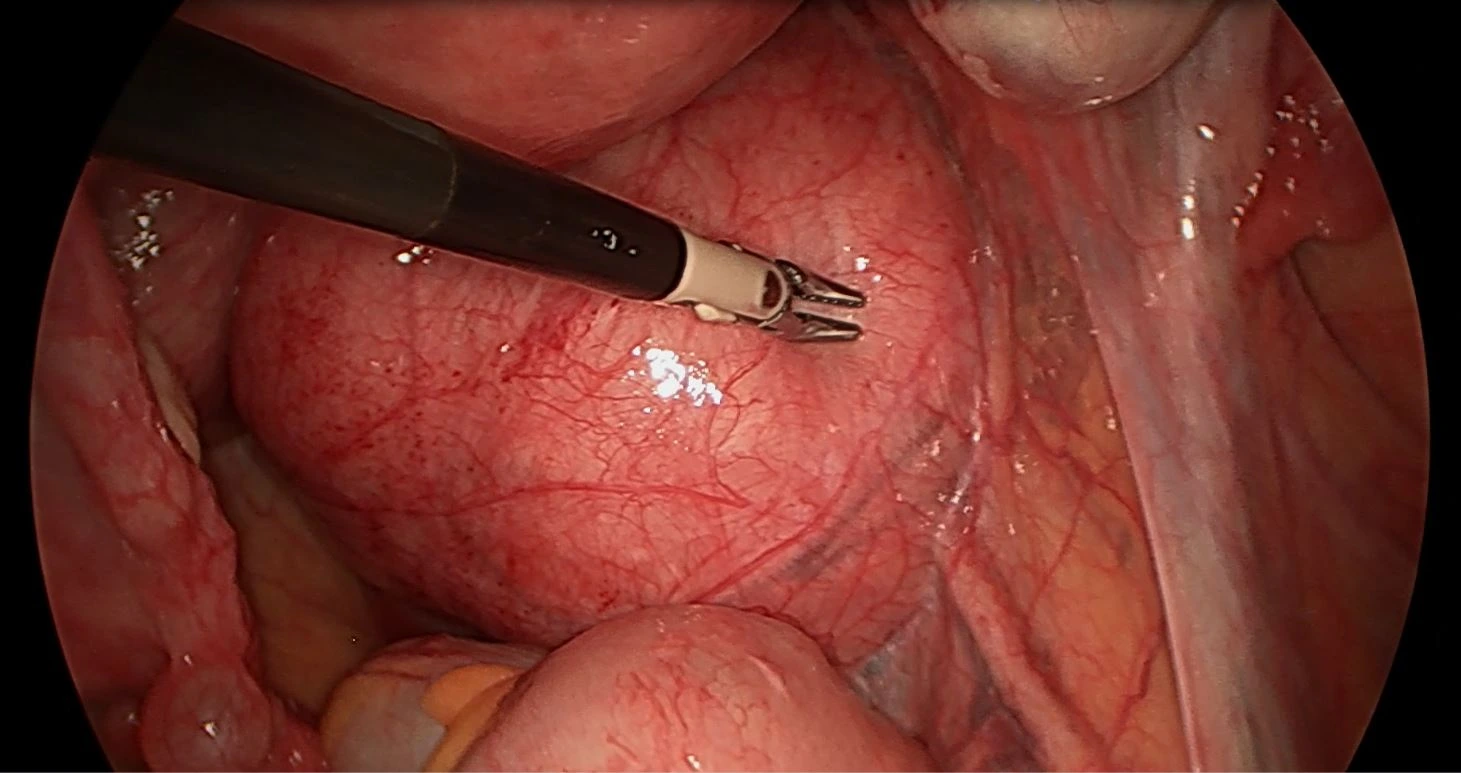
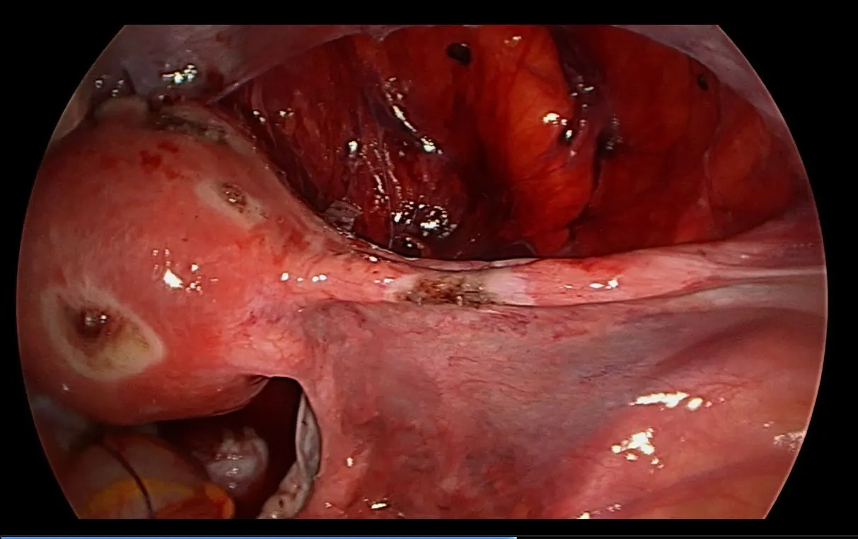
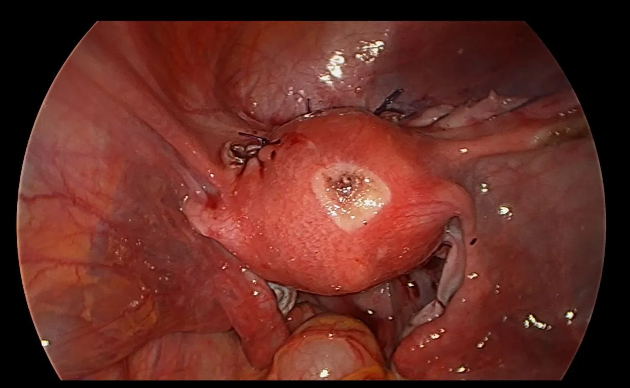
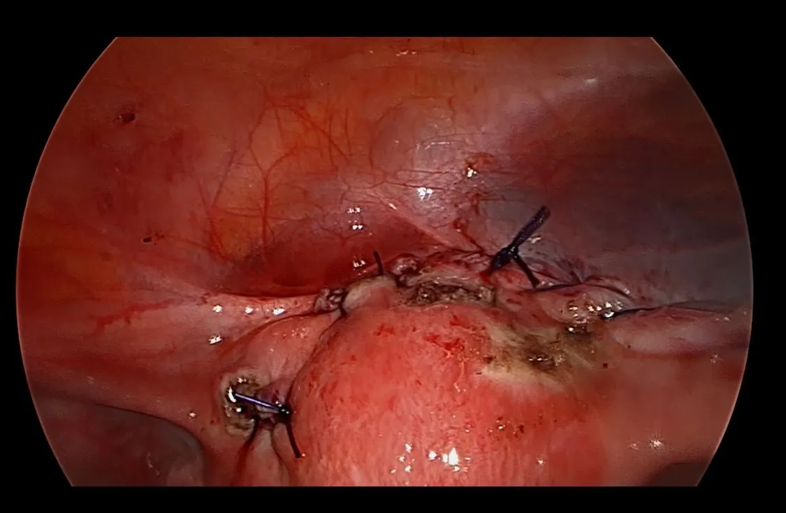
Laparoscopic myomectomy and excision of bilateral ovarian endometriomas


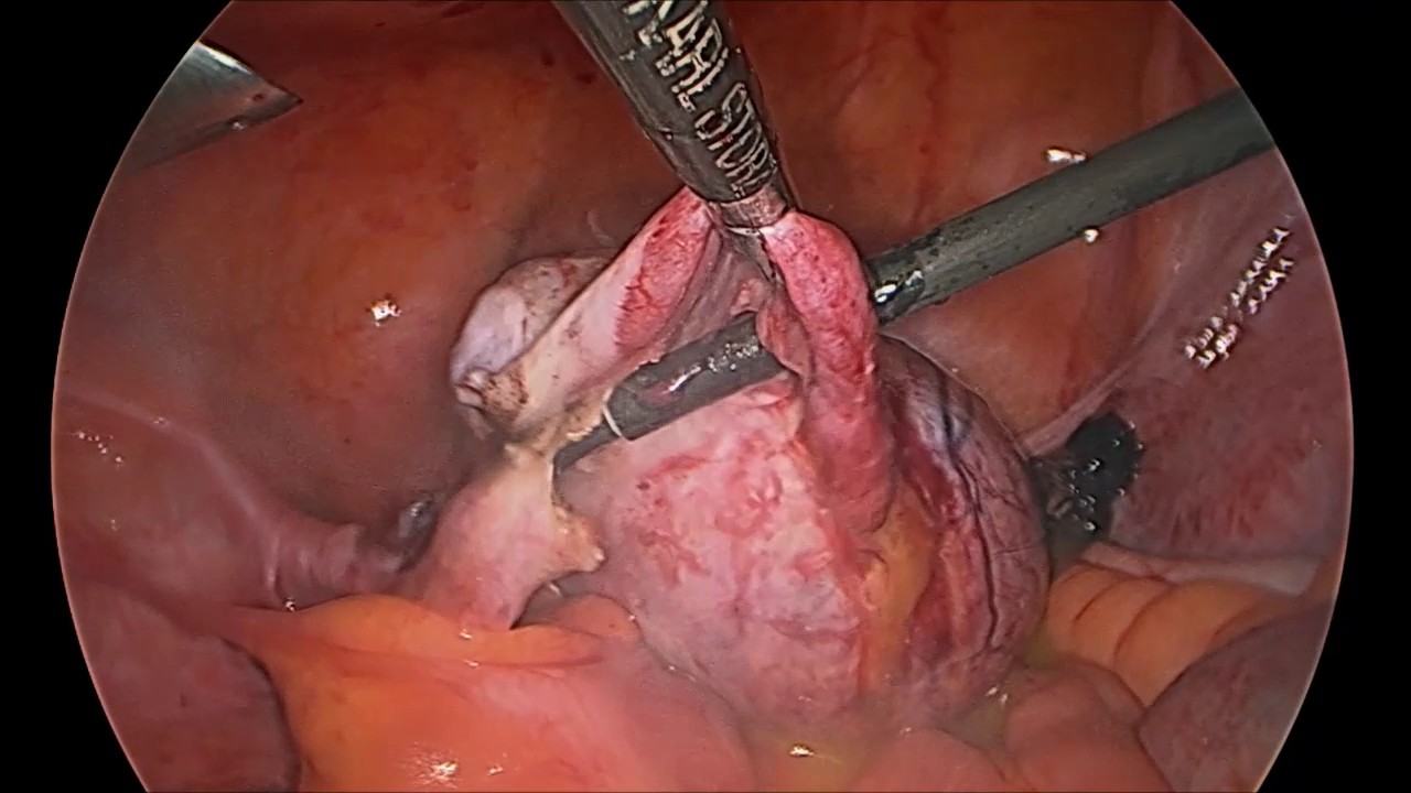
Combined laparoscopic intervention: Atraumatic enucleation of an intramural-subserosal uterine fibroid with simultaneous organ-sparing cystectomy for bilateral ovarian endometriomas, preserving follicular reserve.


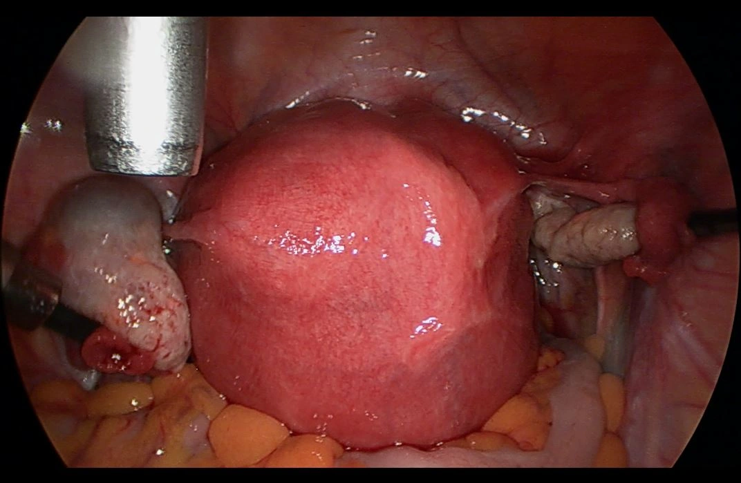
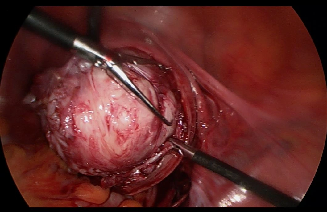
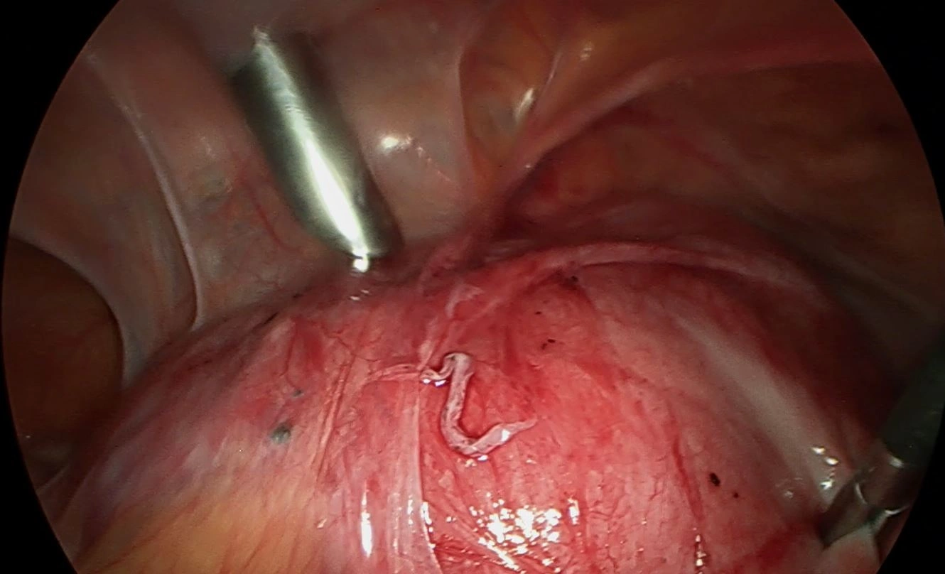
Laparoscopic myomectomy with temporary uterine artery clipping


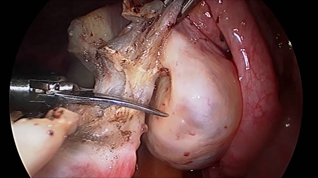
Transient vascular occlusion of the ascending uterine arteries using titanium clips. This achieves a virtually bloodless operative field, minimizes blood loss, and allows anatomical multilayered reconstruction of the myometrium.


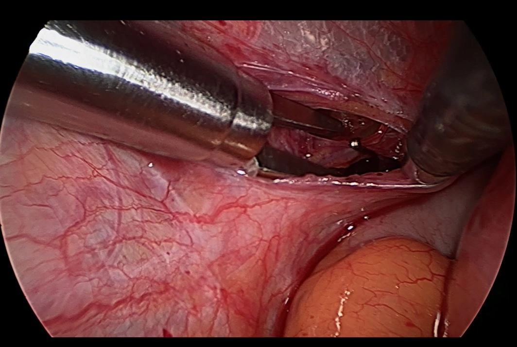
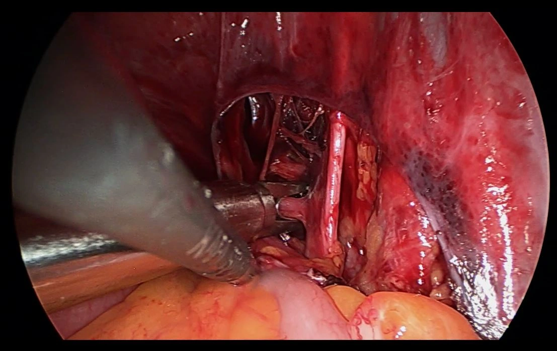
Step-by-step photoprotocol: myomectomy with uterine artery clipping


Surgical sequence: Exposure of the uterine neurovascular bundle, application of a clip to the left uterine artery, hydrodissection of the pseudocapsule, bloodless enucleation, two-layer continuous suturing of the myometrium, and post-revascularization hemostasis.

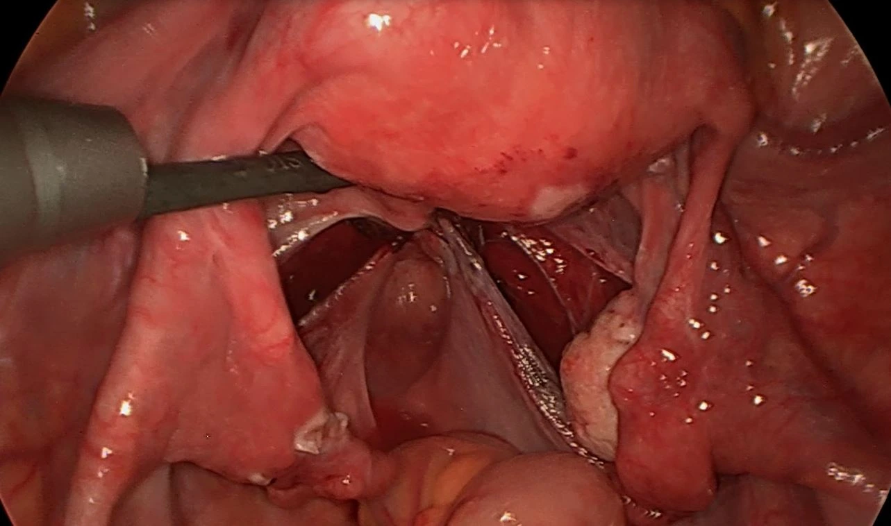
Clipping step 1 Clipping step 2 Clipping step 3 Clipping step 4 Clipping step 5 Clipping step 6 Clipping step 7 Clipping step 8

II. Atypical localizations: retroperitoneal and parametrial fibroids

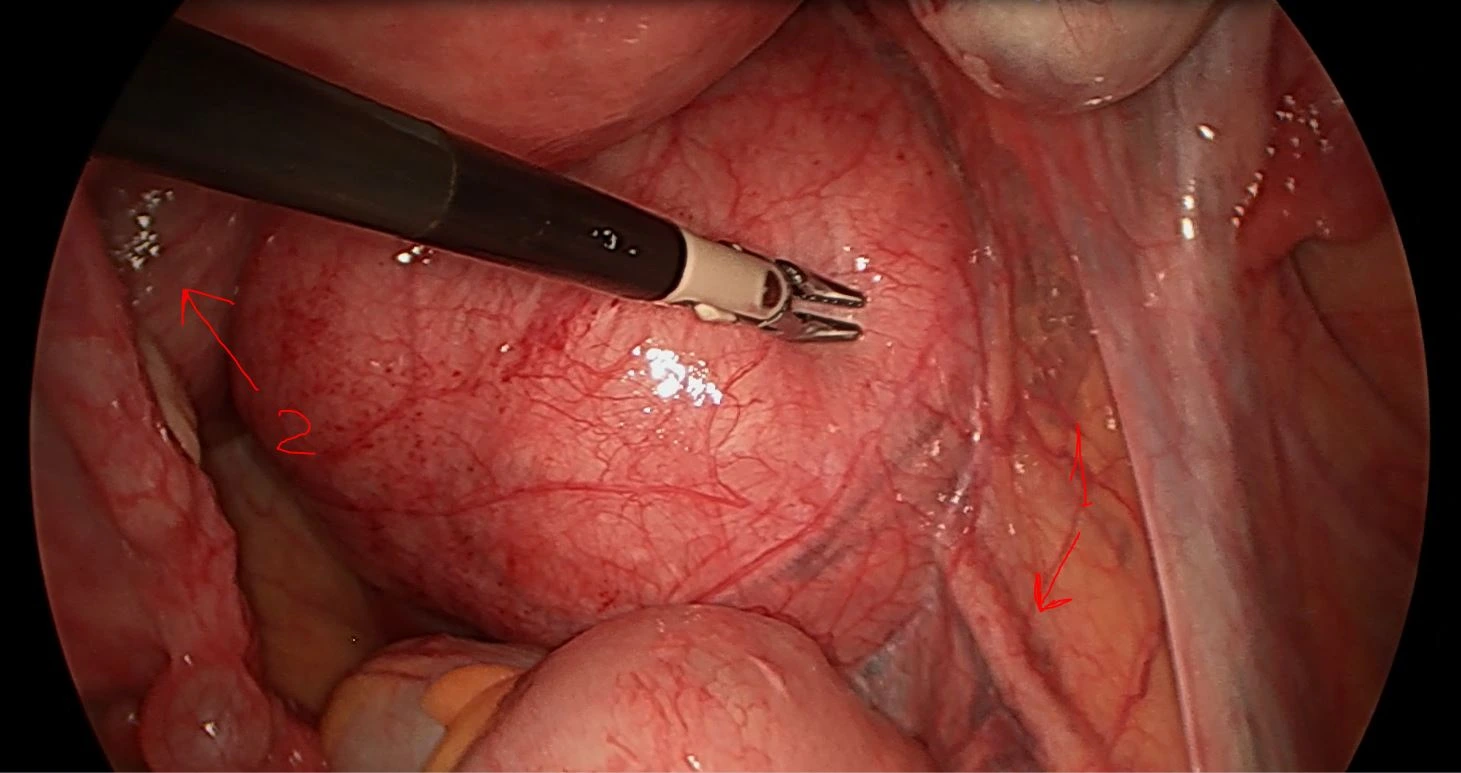
Ureteral compression by a retroperitoneal fibroid with bilateral hydronephrosis


Complex surgical anatomy: Retroperitoneal expansion of the fibroid caused bilateral ureteral obstruction and secondary hydronephrosis. In photo 2-1, arrows indicate the ureteral pathway within the compression zone. Laparoscopic ureterolysis and tumor removal fully restored renal drainage.

Ureterolysis 1 Ureterolysis 2 Ureteral course indicated with arrows Ureterolysis 3 Ureterolysis 4 Ureterolysis 5 Ureterolysis 6

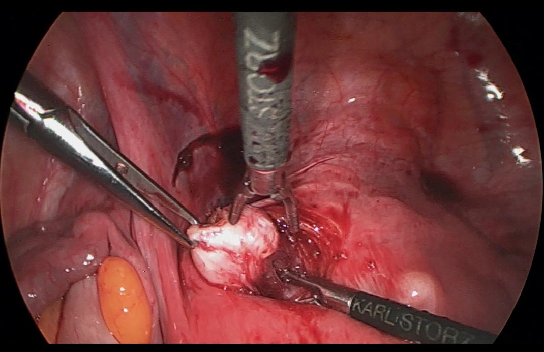
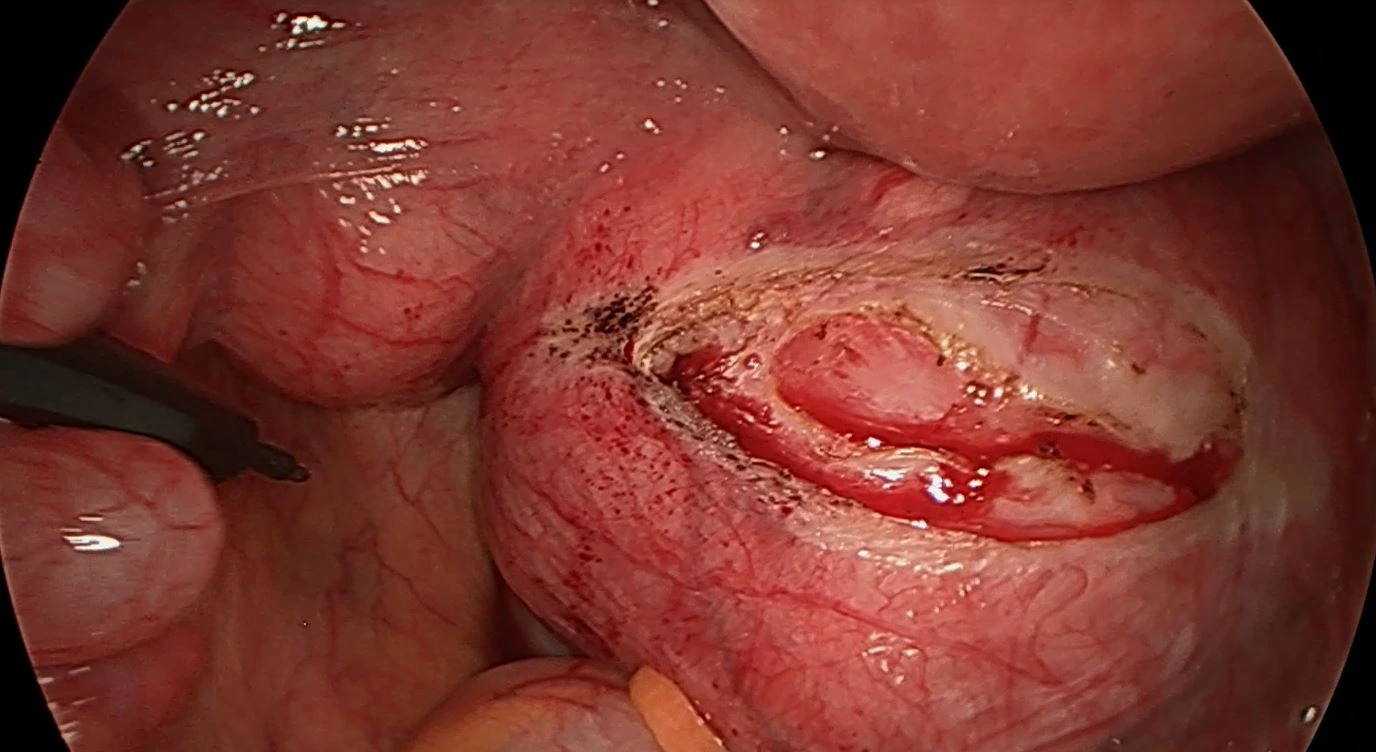
Parametrial (broad ligament) uterine leiomyoma


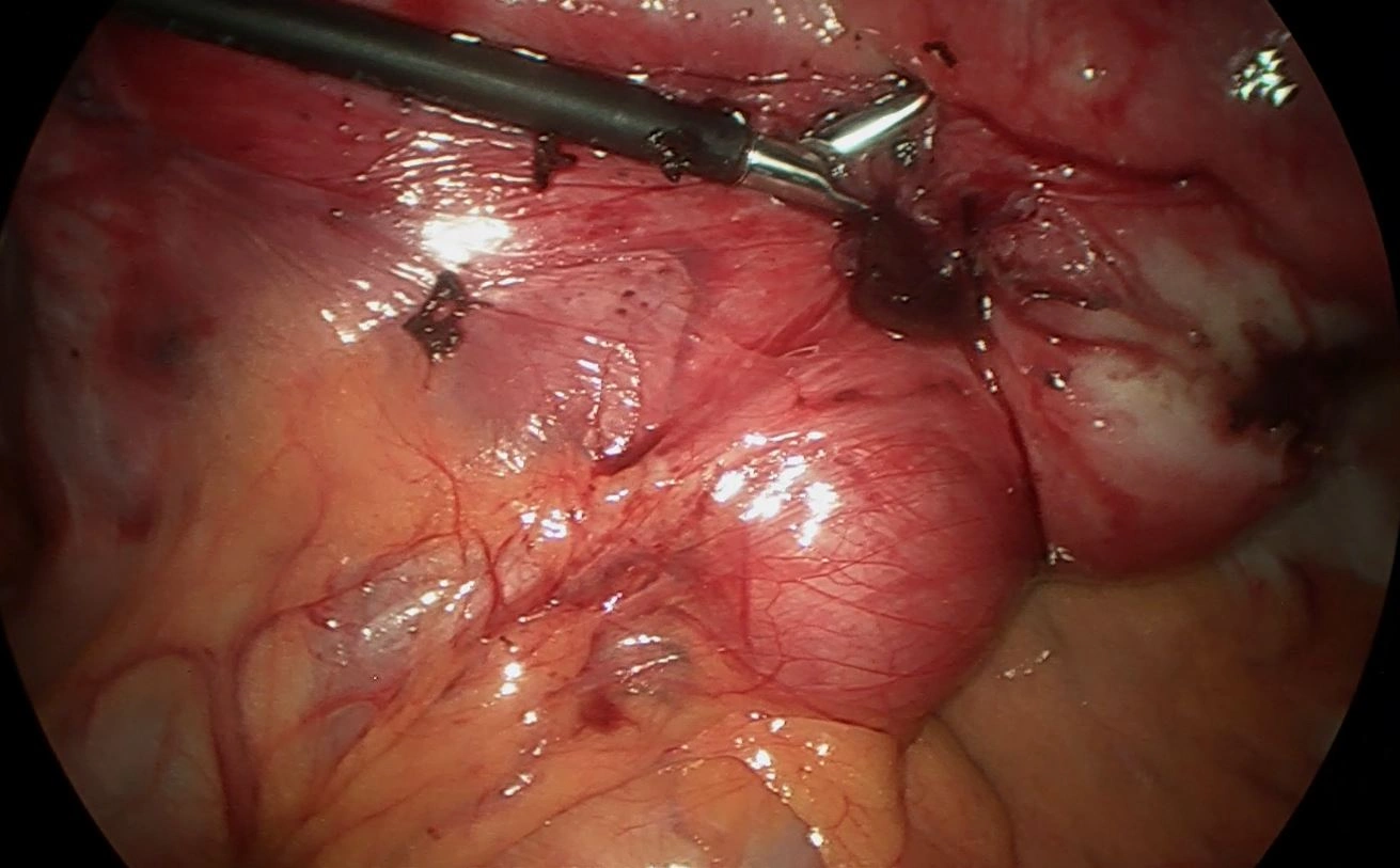
Technically intricate case: The tumor is situated intraligamentously between the leaves of the broad ligament, adjacent to the iliac vessels and ureter. Demands high surgical precision to protect pelvic neurovascular bundles.

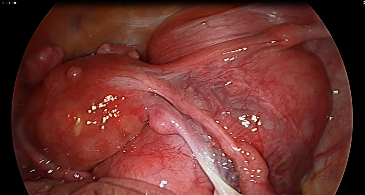
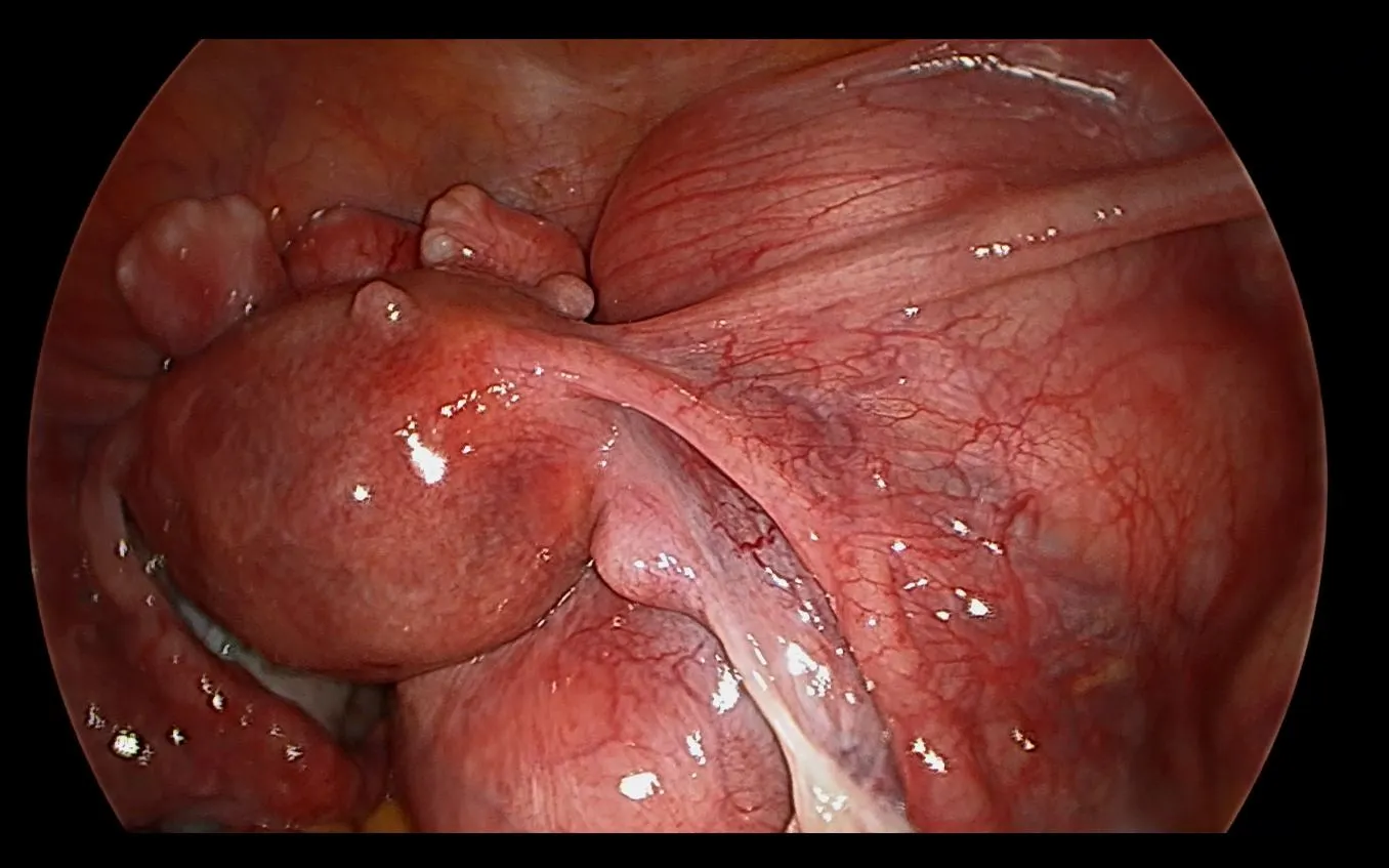
Parametrial fibroid 1 Parametrial fibroid 2 Parametrial fibroid 3 Parametrial fibroid 5 Parametrial fibroid 6 Parametrial fibroid 7

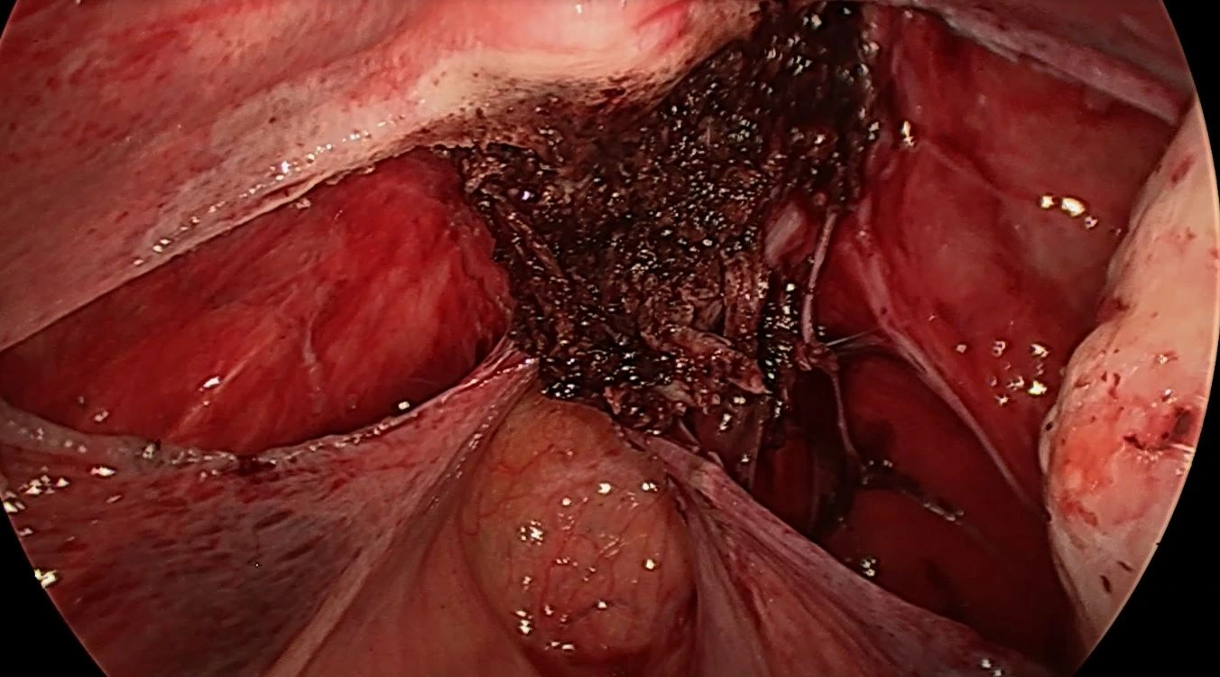
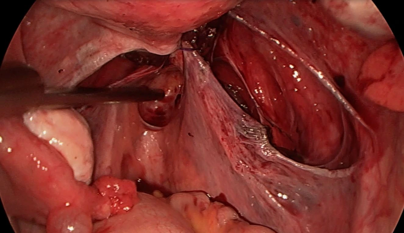
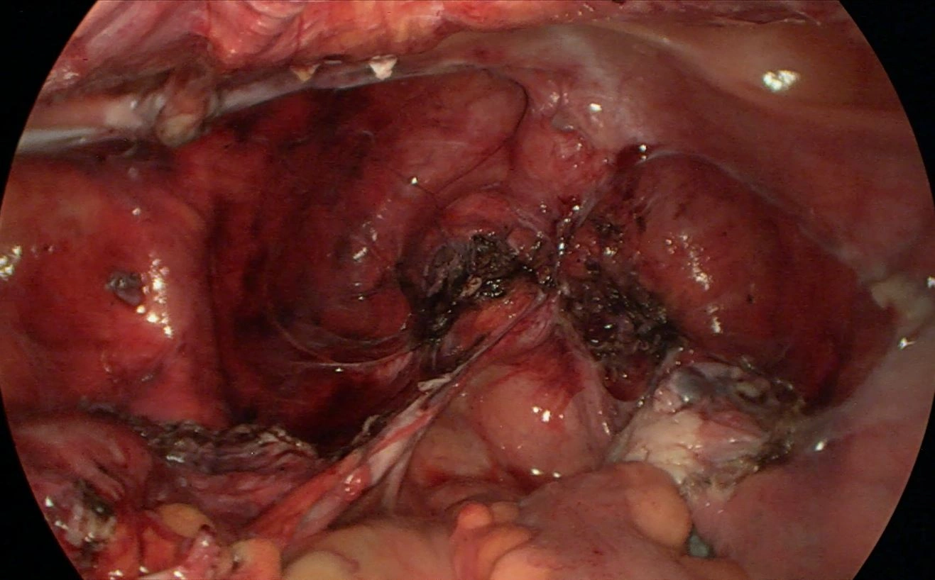
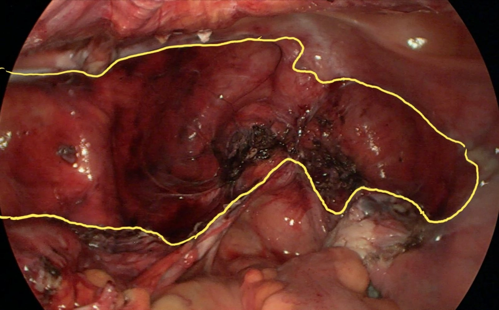
Enucleation of a deep parametrial node with retroperitoneal bed mobilization


Laparoscopic dissection of a deep parametrial tumor. In photo 5, the yellow contour delineates the surgical bed within the retroperitoneal space. Targeted bipolar hemostasis protected autonomic nerve plexuses.

Parametrial node 1 Parametrial node 2 Parametrial node 3 Yellow contour of the tumor bed

III. Giant fibroids: Laparoscopic limits and indications for laparotomy

Year: 2018 Date: 18.01  Laparoscopic removal of a giant uterine leiomyoma


Laparoscopic removal of a giant uterine leiomyoma. All surgical steps are preserved. At 6 min. 35 sec., transection of the left uterine artery leads to acute bleeding; systematic prior vessel skeletonization provided direct exposure for immediate ligation and secure hemostasis.


Year: 2019 Date: 09.04  Laparoscopic hysterectomy for a large uterus (Teamwork)


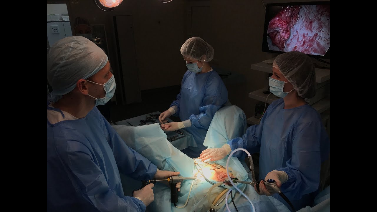
Seamless surgical teamwork during laparoscopic removal of a heavily enlarged multinodular uterus, minimizing anesthesia time and maximizing patient safety.


Morcelloma prevention: In-bag containment morcellation


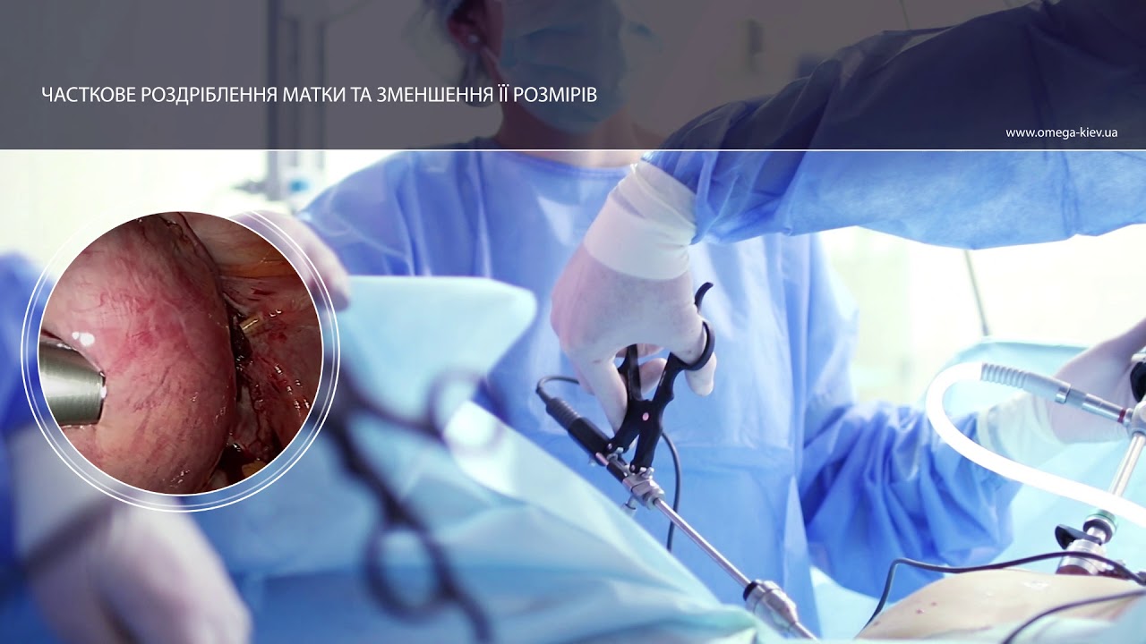
Enclosed electromechanical morcellation within a containment bag. This technique prevents cellular dissemination across the peritoneal cavity, eliminating the risk of iatrogenic parasitic myomas (morcellomas).


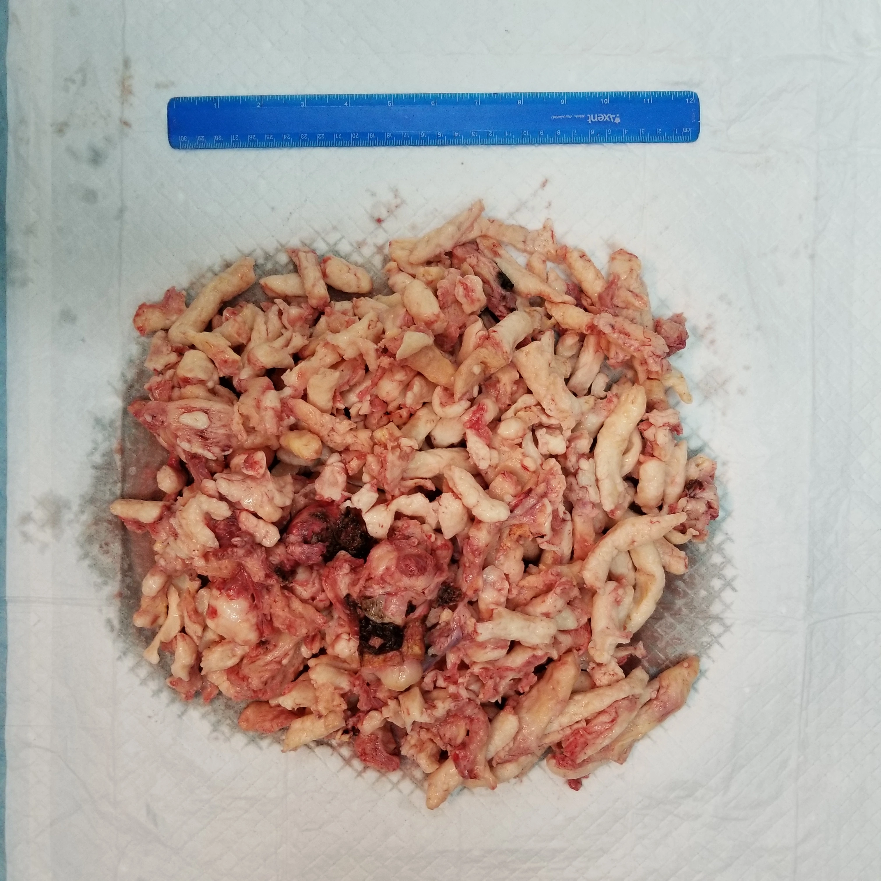
Laparoscopic removal of a giant uterus: morcellated specimen


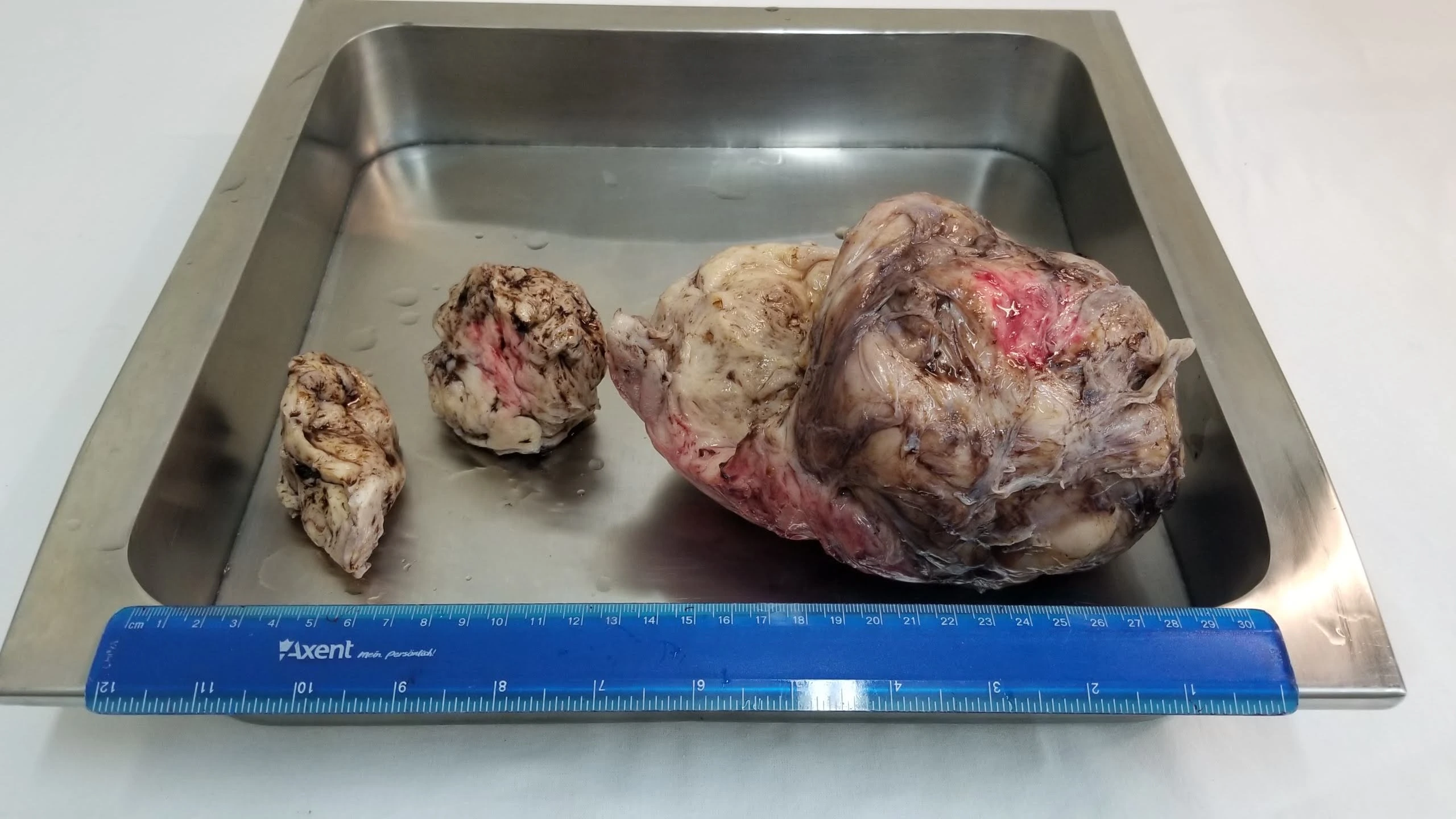
Feasibility of laparoscopic surgery: Giant uteri can be successfully removed through 10–12 mm incisions using containment systems. The photo shows the morcellated specimen alongside a surgical ruler.

Morcellated specimen

Laparotomy for extensive multinodular fibroids (Limits of laparoscopy)


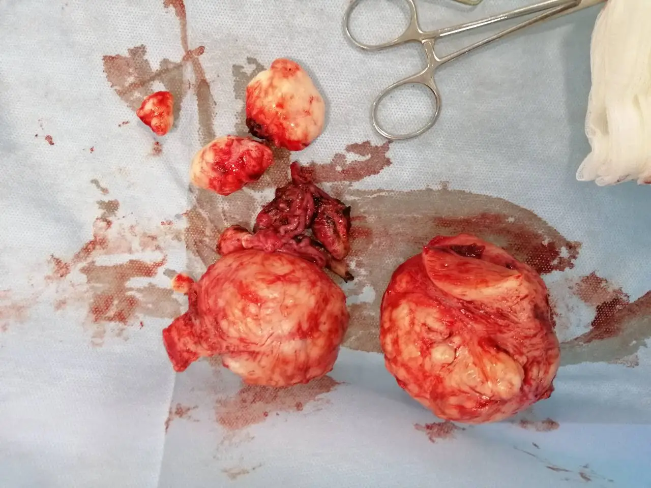
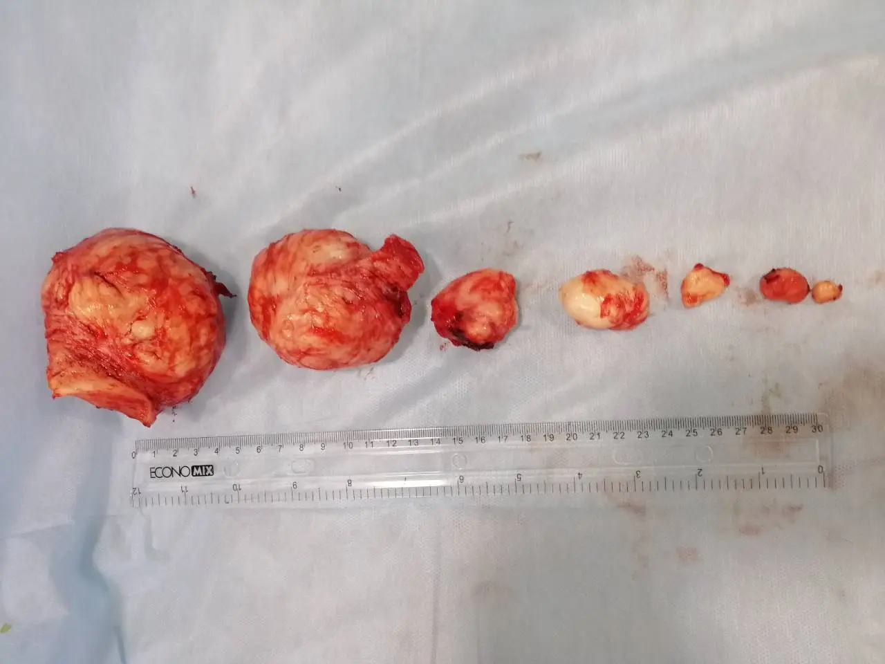
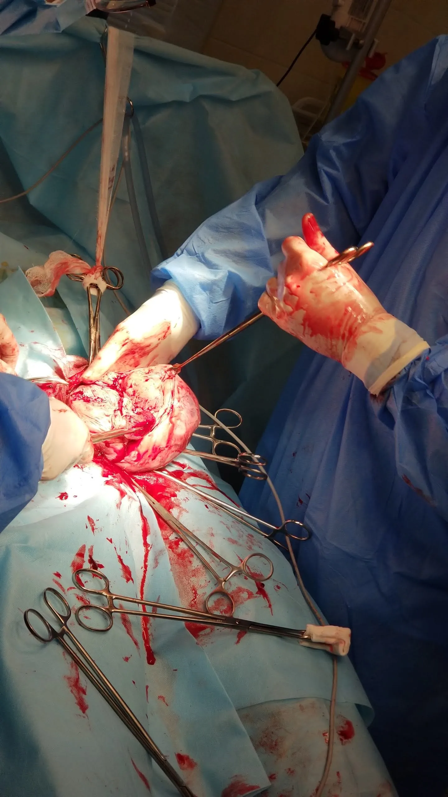
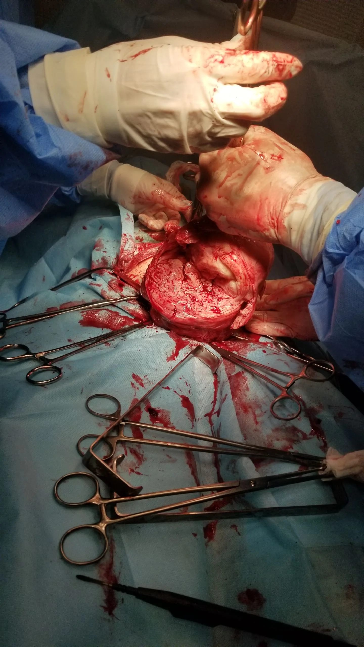
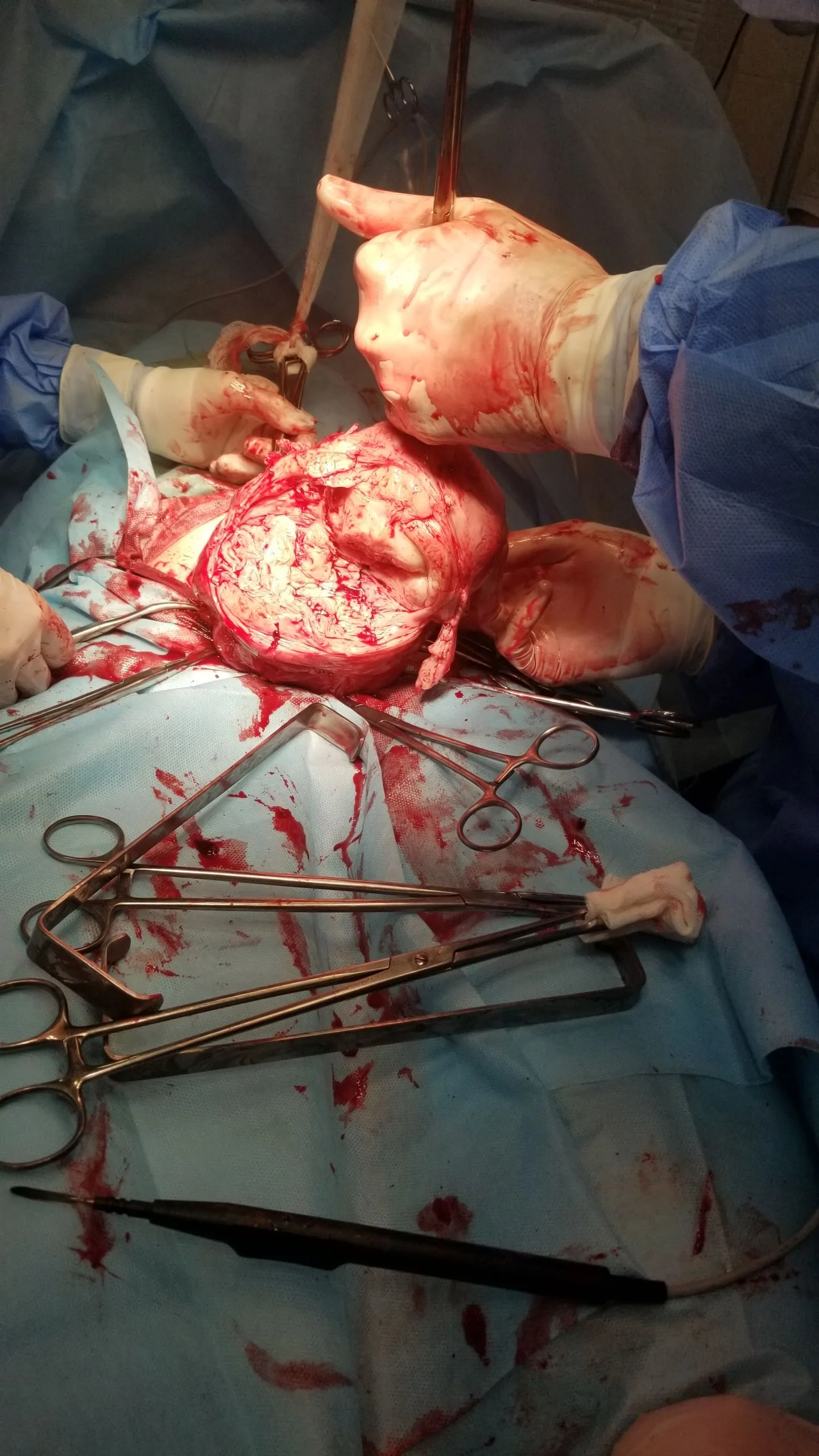
In cases with dozens of deep intramural fibroids throughout the myometrium, laparoscopy carries the risk of leaving occult nodules. Open laparotomy enables full bimanual palpation and resection of all tumors (over 8 dominant fibroids removed) with complete uterine reconstruction.

Laparotomy step 1 Laparotomy step 3 Laparotomy step 4 Laparotomy step 11 Laparotomy step 16 Laparotomy step 22 Laparotomy step 24 Removed fibroids with ruler

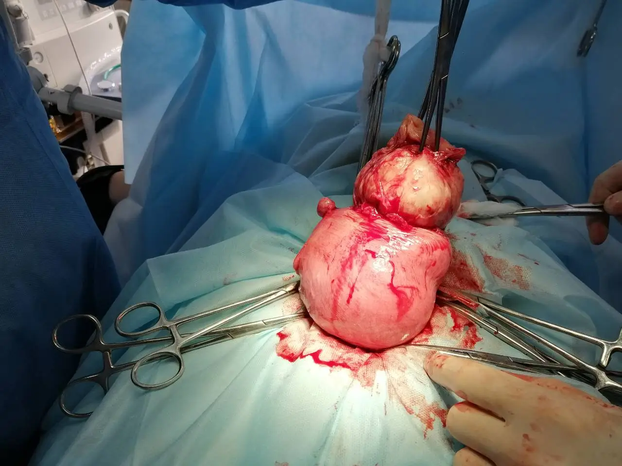
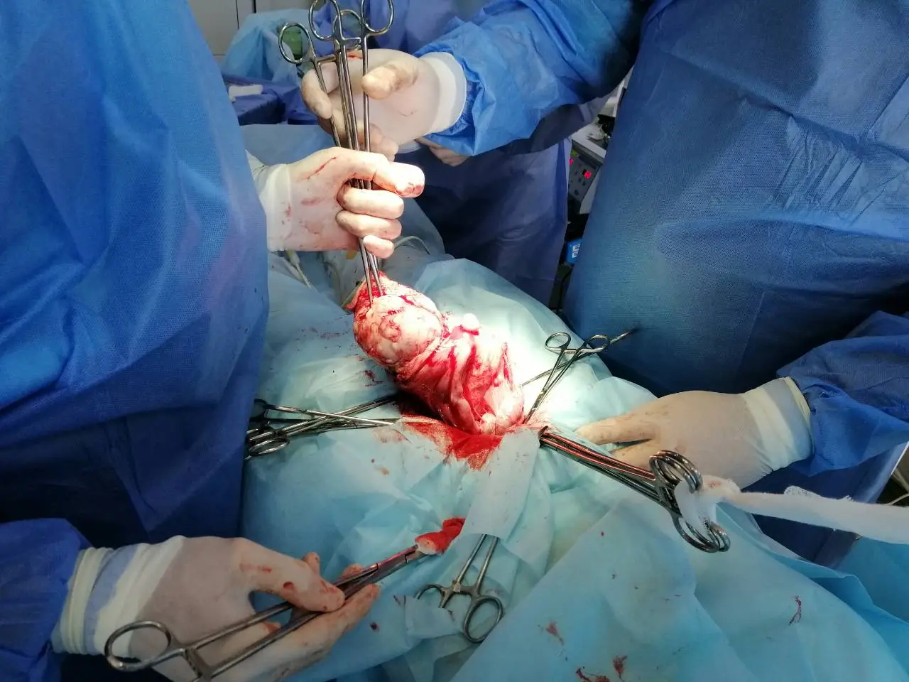
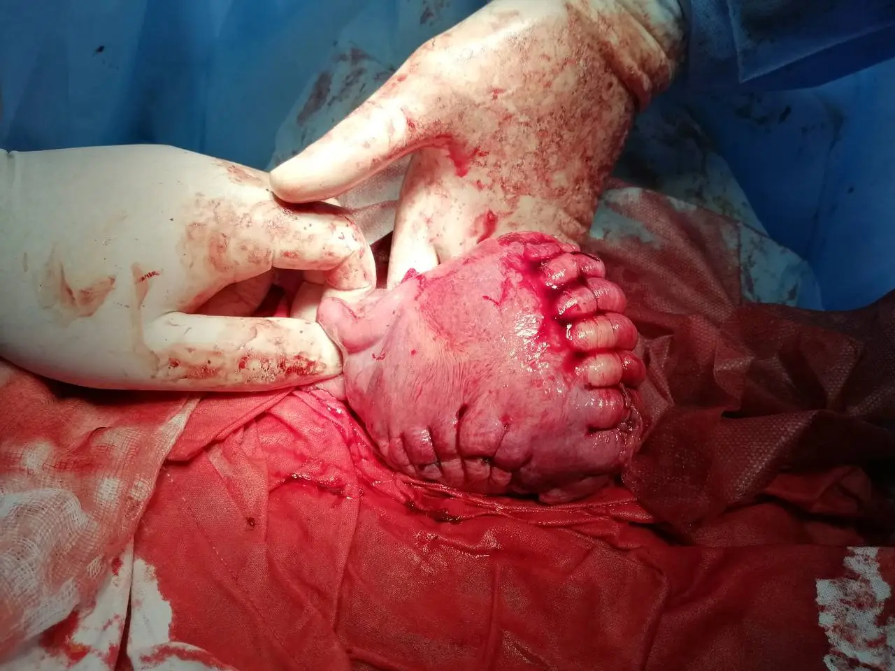
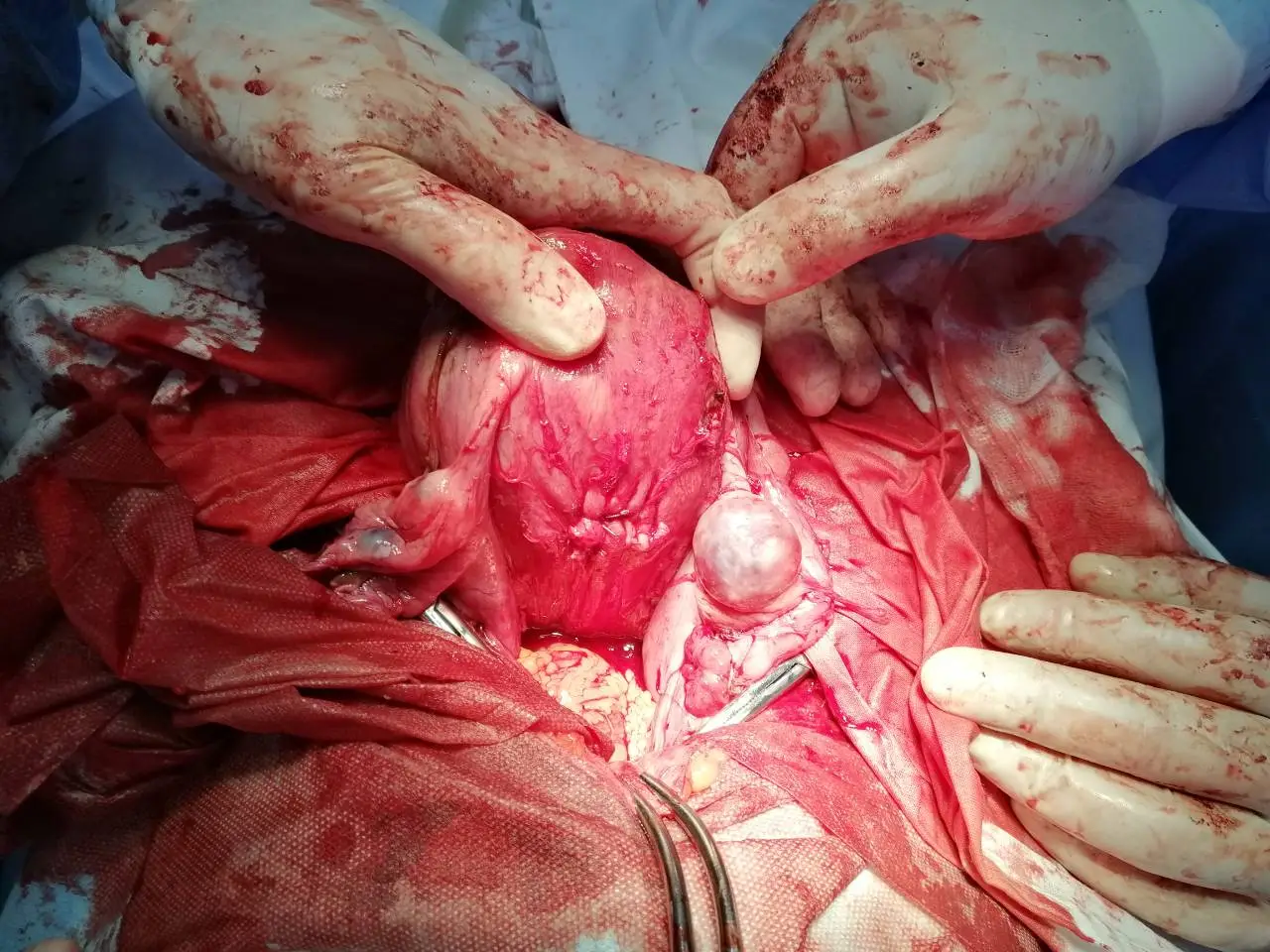
Open surgical resection of a giant abdominal myoma reaching the epigastrium


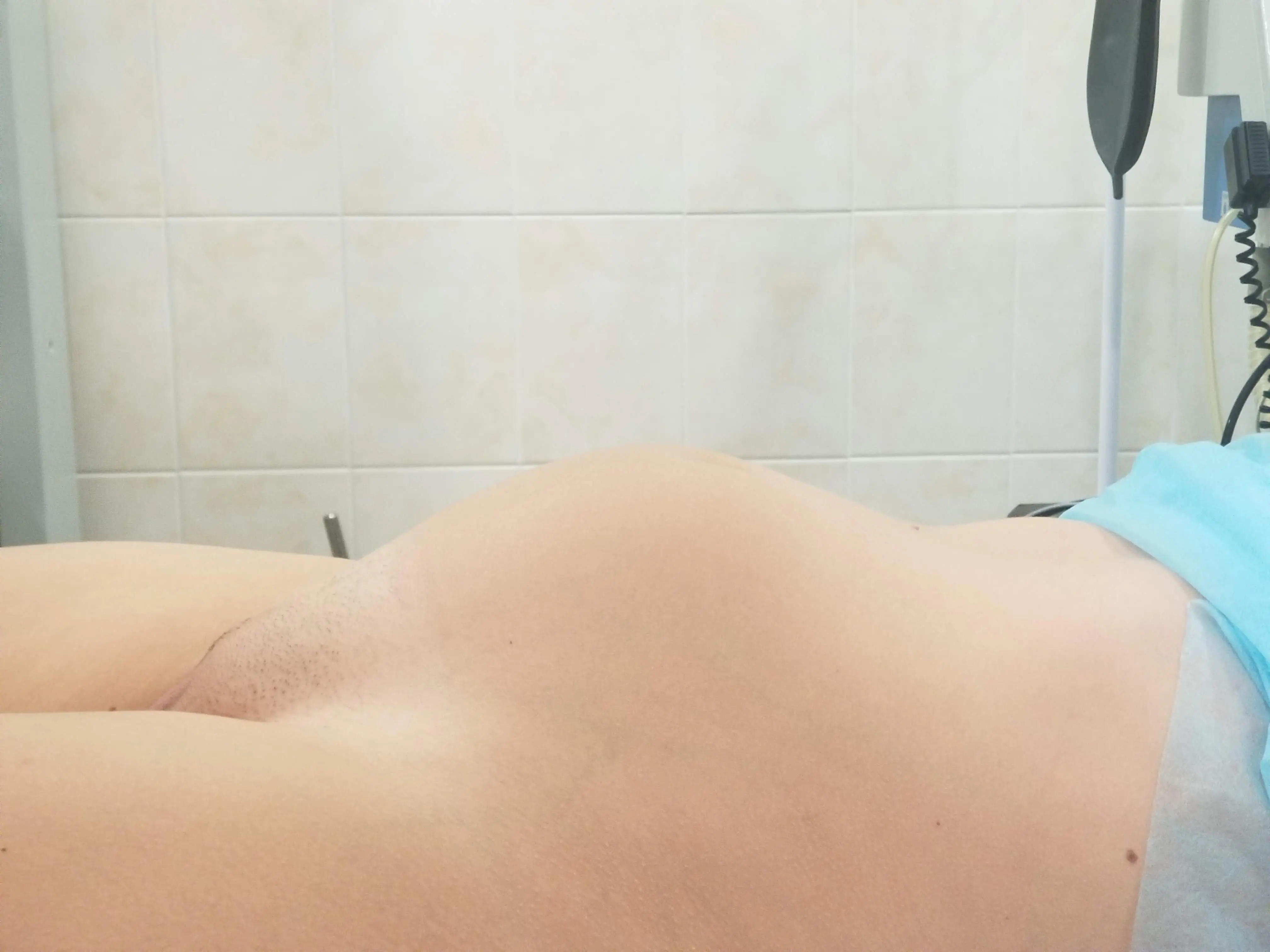
Massive abdominal mass filling the entire pelvic and abdominal cavity up to the epigastric region. Midline laparotomy performed to mobilize and resect the multi-kilogram tumor mass.

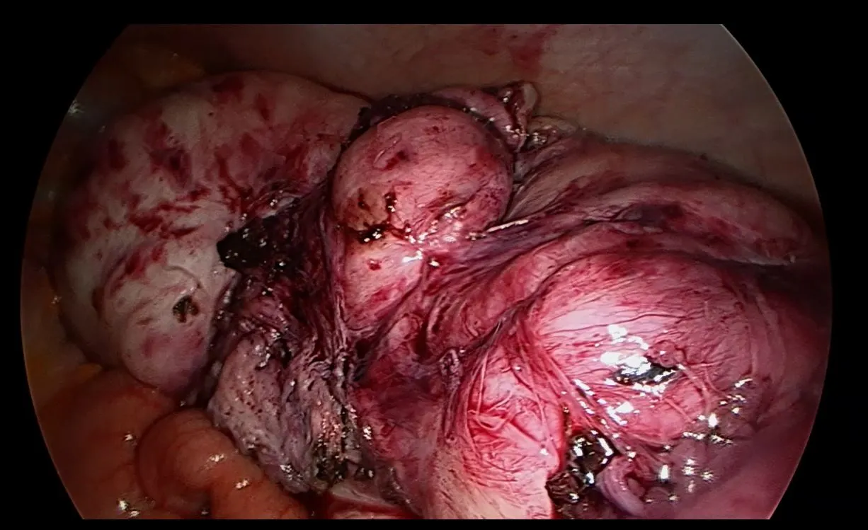
Preoperative view 1 Preoperative view 2 Intraoperative situs Tumor mobilization Resection phase Specimen delivery Gross pathology

IV. Radical procedures: Subtotal (LASH) and total hysterectomy (TLH)


Laparoscopic supracervical / subtotal hysterectomy (LASH)


Laparoscopic removal of the uterine corpus preserving the cervix and pelvic floor suspensory ligaments. Standard choice for patients with normal cervical cytology who have completed family planning.


Total laparoscopic hysterectomy with intrafascial technique (TLH)


Complete removal of the uterus within the endopelvic fascial sheath. Provides precise vascular control, preserves uterosacral ligaments, and protects autonomic nerve pathways against pelvic organ prolapse.