


The main conditions treated by the doctor
Pelvic organ prolapse (POP) and vaginal wall descent represent one of the most common disorders of the female pelvic floor. Among elective surgical interventions in gynecology, genital prolapse ranks third, preceded only by benign tumors and endometriosis. Nearly every second woman over 50 years of age experiences this condition.
A significant subgroup comprises patients with vaginal vault or cervical stump prolapse following radical uterine surgery (hysterectomy or subtotal hysterectomy).
In 85.5% of patients with genital prolapse, comorbid functional disorders of adjacent pelvic organs develop:
• urinary incontinence — in 70.1% of patients;
• defecatory dysfunction — in 36.5%;
• pain during sexual intercourse (dyspareunia) — in 53.3% of patients.
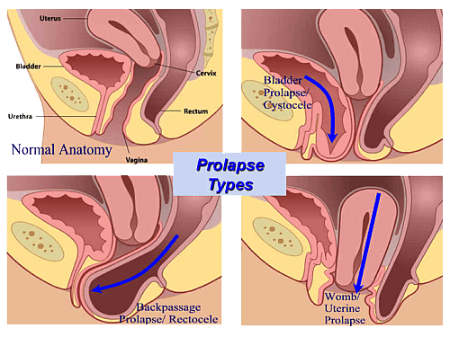
The prevalence of genital prolapse and related urinary dysfunction increases steadily with age.
Urinary incontinence is observed in up to 30% of women under 55 years and in more than 75% of women over 70 years of age.
Classification
Malinowski's clinical classification is widely recognized for its simplicity. It distinguishes three degrees of prolapse (Fig. 1):
• Grade I — the cervix descends to the vaginal introitus but does not protrude beyond it;
• Grade II — the cervix extends beyond the introitus, while the uterine corpus remains above it (incomplete prolapse);
• Grade III — complete uterine prolapse (the entire uterus resides outside the vaginal introitus).
Fig. 1:
urinary bladder — a
normally positioned uterus — b
vagina — c
vaginal entrance — d
uterine cervix — e
incorrect position of the uterus (3 degrees of prolapse) — f
In contemporary clinical practice, the standardized international POP-Q (Pelvic Organ Prolapse Quantification) system is employed. It quantitatively assesses anatomical support using nine specific anatomical landmarks:
Aa — anterior vaginal wall (urethrovesical junction);
Ba — most dependent point of upper anterior vaginal wall;
Ap — lower posterior vaginal wall;
Bp — upper posterior vaginal wall;
C — cervix or vaginal cuff;
D — posterior fornix / pouch of Douglas;
TVL — total vaginal length;
GH — genital hiatus;
PB — perineal body.
According to the POP-Q staging system:
Stage 0 — no prolapse. Points Aa, Ap, Ba, Bp are all at -3 cm; points C and D reside at normal baseline values.
Stage I — the most distal portion of the prolapse is > 1 cm above the hymenal ring (value < -1 cm).
Stage II — the leading edge is between 1 cm above and 1 cm below the hymenal plane (-1 cm to +1 cm).
Stage III — the leading edge extends > 1 cm beyond the hymen, but does not exceed (TVL - 2) cm.
Stage IV — complete eversion/procidentia. The most distal part extends to within (TVL - 2) cm of the total vaginal length.
Etiology and Risk Factors
The primary underlying mechanism is pelvic floor weakness and failure of the supporting fascial and muscular apparatus to sustain pelvic visceral load. Genital prolapse represents a true pelvic floor hernia. The condition arises from multifactorial causes, where vaginal parity is an essential, though not exclusive, predisposing factor.
Key risk factors include:
• obstetric trauma (fetal macrosomia, prolonged or operative vaginal delivery, perineal tears);
• systemic connective tissue weakness (undifferentiated connective tissue dysplasia, recurrent hernias);
• hypoestrogenism (menopause, impaired steroidogenesis);
• chronic intra-abdominal hypertension (chronic cough, constipation, strenuous physical labor).
Clinical Presentation
Major clinical manifestations of pelvic organ prolapse:
• widening/gaping of the genital hiatus at rest or during Valsalva maneuver;
• visible or palpable bulging of vaginal walls / cervix beyond the introitus;
• sensation of a pelvic foreign body or heaviness;
• perineal discomfort and difficulty during prolonged walking;
• dragging lower abdominal, sacral, and lumbar pain;
• dyspareunia (pain during intercourse);
• postcoital spotting or bleeding.
Descent of the uterus and pelvic organs leads to pelvic venous stasis, exacerbating persistent dragging pain and pelvic fullness that characteristically worsen towards the end of the day or after physical exertion.
Chronic friction and mucosal desiccation induce trophic alterations, erosions, and decubitus ulcerations, often accompanied by contact bleeding. In postmenopausal women, severe vaginal atrophy further degrades natural anti-infective barriers, causing recurrent vaginitis.
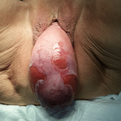
A key feature of genital prolapse is the associated dysfunction of adjacent pelvic organs. Urological symptoms range from stress urinary incontinence and pollakiuria to obstructive voiding difficulties and acute urinary retention in advanced prolapse stages. Advanced pelvic floor defects also frequently manifest as rectocele-induced defecatory dysfunction and fecal/gas incontinence.