


The main conditions treated by the doctor
Among the pathological processes of the uterine cavity, the most common are: endometrial polyps, endometrial hyperplasia, submucosal uterine leiomyoma, adenomyosis (internal endometriosis), and intrauterine synechiae.
Endometrial hyperplasia is a pathological proliferation of the uterine mucosa with predominant involvement of glandular structures.
According to the WHO classification, endometrial hyperplastic processes include hyperplasia, polyps, and atypical changes in the endometrial mucosa.
Uterine polyps usually develop due to the proliferation of the basal layer glands of the endometrium. Initially, glandular growth and surrounding stroma form a polypoid outgrowth on a broad base; subsequently, a stalk of varying thickness develops, which may contain vascular and smooth muscle structures.
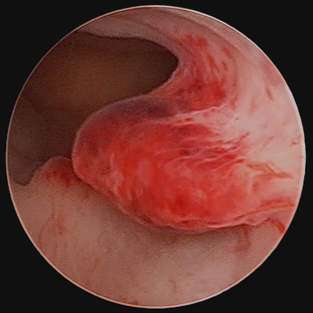
Polyps can cause infertility. The primary clinical concern is the risk of malignant transformation.
Clinically, polyps manifest as intermenstrual spotting, abnormal uterine bleeding, or they may remain entirely asymptomatic.
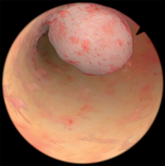
The diagnosis of a uterine polyp is primarily established by transvaginal ultrasound. Polyps may be single or multiple, most frequently localized in the uterine fundus or tubal cornua.
Upon initial detection, a follow-up ultrasound is recommended right after the subsequent menstrual cycle. If the formation persists, hysteroscopic removal is indicated.
For smaller polyps (up to 2–2.5 cm), removal using an ultrathin mini-hysteroscope is possible, requiring no cervical dilation. This minimally invasive method is especially valuable for nulliparous women, as it prevents cervical trauma and fully preserves reproductive potential.
For large polyps (exceeding 3 cm), submucosal fibroids, or dense intrauterine synechiae, a bipolar hysteroresectoscope is utilized.
Unlike classic monopolar systems, this instrument operates in isotonic saline solution, significantly improving surgical safety, allowing extended operative time, and enabling the precise resection of larger intracavitary formations (up to 5–6 cm).